OVERVIEW

The key question isn’t “present or absent.”

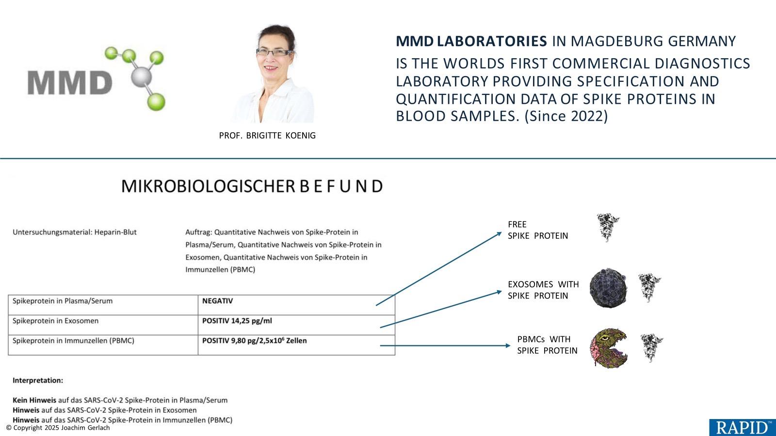
The more useful question is: where is spike located — and what form is it in? Different methods see different compartments, which changes interpretation.

Clinical framing: results can differ not only because of sensitivity, but because sample type and analytical target differ.
What can vary
  • Compartment (serum, exosomes, PBMCs)
  • Molecular form (intact vs fragments)
Why it matters
  • A “negative” result may not rule out other compartments
  • Method choice affects clinical confidence
METHODS

Three ways to look for spike

Each method answers a different clinical question: screen, measure, or confirm.

Lateral Flow (Antigen-Release)
Screen Fast Semi-quantitative
Best for: rapid triage / first-look screening.
Output: signal strength (not a concentration).
ELISA
Measure Quantitative Monitoring
Best for: baseline + trend monitoring over time.
Output: numeric result (assay-dependent).
Mass Spectrometry (MS)
Confirm Peptide-level Sequence-confirmed
Best for: complex cases and deeper resolution.
Output: peptide detection with molecular confirmation.
Rule of thumb: use the simplest method that answers the question — then escalate when compartment access or peptide-level confirmation matters.

Compartment mapping

What You Learn Depends on Where You Look

A “negative” result can mean different things depending on the compartment tested.

Compartment
What it suggests
Tests that can access it
Serum / Plasma
Circulating accessible antigen
Lateral Flow, ELISA, MS
Exosomes
Vesicle-packaged antigen
ELISA (with isolation), MS
PBMCs
Intracellular immune-cell-associated antigen
MS, Lateral Flow (with lysis)
Whole Blood
Integrated signal
Lateral Flow

Testing Method

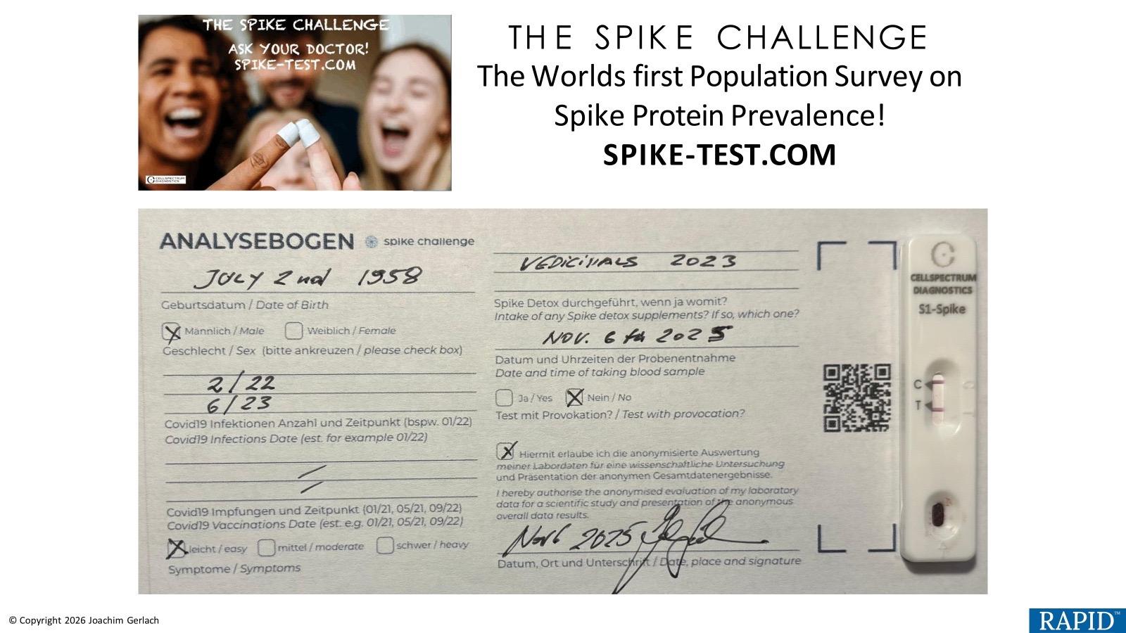
Lateral Flow (Antigen-Release)

A rapid screening approach designed to answer one question quickly: is there a detectable spike-antigen signal?

Fast / Point-of-care Semi-quantitative Broad blood coverage (release/lysis)

What it measures

  • Spike antigen signal after an antigen-release / lysis step.
  • Can access blood compartments that may include free, exosomal, and cell-associated antigen signals depending on format/prep.

Best used for

  • Rapid triage / screening workflows
  • “Screen first” decision making before more involved testing
  • Practice pilots where speed matters

Output

Semi-quantitative (band intensity / signal strength), not a precise concentration value.

How to interpret it

If positive

Treat as a screening signal. Consider confirmation/quantification with ELISA or MS based on clinical complexity.

If negative

A negative screen does not necessarily exclude other compartments/forms. If suspicion persists, consider deeper methods (MS) and/or compartment-specific approaches.

Why results can differ from ELISA or MS

Different tests “see” different biological targets and compartments. Antigen-release formats aim to broaden what becomes detectable in a whole-blood workflow, while ELISA depends on antibody-accessible epitopes and MS detects peptide fragments after digestion.

When to move beyond screening

If symptoms are persistent/complex or you need molecular confirmation, step up to MS; if you want a numeric value for accessible circulating antigen, consider ELISA.

Educational use only. This page summarizes analytical methodologies and does not replace clinical judgment or constitute diagnostic guidance.

Testing Method

ELISA (Enzyme-Linked Immunosorbent Assay)

A quantitative immunoassay designed to measure antibody-accessible spike antigen — useful for baseline and trend monitoring.

Quantitative Accessible + scalable Monitoring-friendly

What it measures

  • Spike antigen that retains the relevant epitope(s) for antibody binding.
  • Most commonly: free spike in plasma/serum.
  • Can be extended to exosomes or PBMCs with added isolation/lysis steps (prep-dependent).

Best used for

  • Mild/early presentations (baseline)
  • Cost-sensitive screening
  • Trend monitoring over time (repeatable)

Output

Numeric concentration (assay-dependent ranges), supporting trending and comparison within the same methodology.

Practical workflow

Start with ELISA when…

You want a quantitative baseline and symptoms are mild-to-moderate, or you need a scalable monitoring tool.

Escalate when…

ELISA is negative/equivocal but symptoms persist, or the presentation suggests deeper compartments/forms — consider MS for peptide-level confirmation.

Why an ELISA result can be “negative” while symptoms persist

ELISA depends on intact antibody-binding epitopes and on the antigen being in an accessible compartment. Fragmented/modified or intracellular forms may not register without additional prep.

Best practice for monitoring

When trending over time, use consistent pre-analytics and the same methodology wherever possible to reduce variability.

Educational use only. ELISA and MS are often best viewed as sequential tools: ELISA for accessible quantification/monitoring, MS for deeper confirmation in complex cases.

Testing Method

Mass Spectrometry (MS)

A peptide-level approach designed for molecular confirmation: which spike peptides are present, and at what level.

Sequence-confirmed Fragment-sensitive Complex cases

What it measures

  • Spike peptides detected after digestion (molecular confirmation).
  • Can be applied to serum/plasma, exosomes, and PBMC/cell-associated compartments (processing-dependent).

Best used for

  • Moderate-to-severe or persistent symptoms
  • When ELISA is negative but suspicion remains
  • When deeper compartment/form insight matters

Output

Quantitative peptide detection with sequence-level confirmation (lab/platform dependent).

Why MS is often the escalation step

Reduces false reassurance

MS can detect peptide fragments that antibody-based assays may miss when epitopes are altered or fragmented.

Supports complex decision making

Used when the clinical picture is disproportionate, multisystem, or persistent and higher resolution is needed.

Why MS and ELISA can disagree

ELISA depends on antibody recognition of specific epitopes; MS detects peptides after digestion, enabling confirmation even when spike is fragmented or modified.

Practical sequencing

A common pathway is ELISA for baseline/monitoring and MS as the escalation method for severity, persistence, or uncertainty.

Educational use only. MS is frequently positioned as a confirmatory method for complex cases and deeper biological compartment questions.

INTERPRETATION

Why Test Results May Differ

It is not uncommon for ELISA and Mass Spectrometry results to differ. This does not necessarily indicate error.

It reflects biology.
Epitope Dependence
ELISA relies on intact epitope recognition. Fragmented or structurally modified spike may evade antibody capture.
Intracellular Reservoirs
Spike stored within PBMCs or immune cells may not be reflected in serum-based ELISA testing unless lysis is performed.
Exosomal Compartment
Vesicle-associated spike requires isolation prior to detection. Standard serum evaluation may underrepresent this compartment.
Peptide-Level Detection
Mass Spectrometry uses digestion + peptide identification, enabling detection beyond antibody-binding limitations.
Key Insight
A negative serum ELISA does not necessarily exclude persistent spike. Biological compartment, molecular form, and analytical methodology determine visibility.
ANALYTICAL DEPTH

Analytical Resolution: How Deep Does Each Test Go?

Screening, quantification, and peptide-level confirmation are different analytical goals. This comparison summarizes what each method is designed to resolve.

Dimension
Lateral Flow
ELISA
Mass Spectrometry
Output Type
Semi-quantitative
Quantitative
Quantitative + sequence-confirmed
Epitope Dependent
Yes
Yes
No (peptide-based)
Fragment Detection
Limited
Limited
Strong
Intracellular Access (PBMC)
Via release/lysis
Limited (prep dependent)
Yes
Best Use
Rapid screening
Quantification + monitoring
Confirmation + complex cases

Practical takeaway: analytical resolution increases from screening (Lateral Flow) to quantification (ELISA) to peptide-level confirmation (Mass Spectrometry).

Workflows

Practical Testing Pathways

Different clinical contexts call for different sequencing. These pathways summarize common decision flows.

A

Stepwise (Cost-Sensitive)

Start broad, then escalate as needed.

  1. Start with ELISA (serum/plasma).
  2. If positive → consider trending/monitoring.
  3. If negative but symptoms persist → escalate to Mass Spectrometry.

B

Direct-to-MS (Clinically Complex)

High-resolution confirmation first.

  1. Begin with Mass Spectrometry in persistent, multisystem, or severe presentations.
  2. Use results to clarify compartment + molecular form.
  3. Use ELISA for longitudinal monitoring when appropriate.
C

Rapid Triage First

Fast signal → confirm with lab method.

  1. Lateral Flow (antigen-release) for rapid screening.
  2. If positive → confirm/quantify with ELISA or MS depending on complexity.
  3. If negative but suspicion remains → consider MS for deeper detection.

Core principle

These Are Not Competing Tests

ELISA and Mass Spectrometry are sequential tools in a rational diagnostic funnel.

ELISA

  • Accessible entry point
  • Quantitative and scalable
  • Best for screening and monitoring trends

Mass Spectrometry

  • High-resolution confirmation
  • Detects peptides/fragments and deeper compartments
  • Best for complex, persistent, or severe cases

Used correctly, these tools reduce diagnostic ambiguity, prevent premature closure, and improve clinical confidence.

Educational use: This page summarizes analytical methodologies used in research and monitoring contexts for spike antigen detection. It is intended for educational purposes and does not replace clinical judgment or constitute a diagnostic guideline.